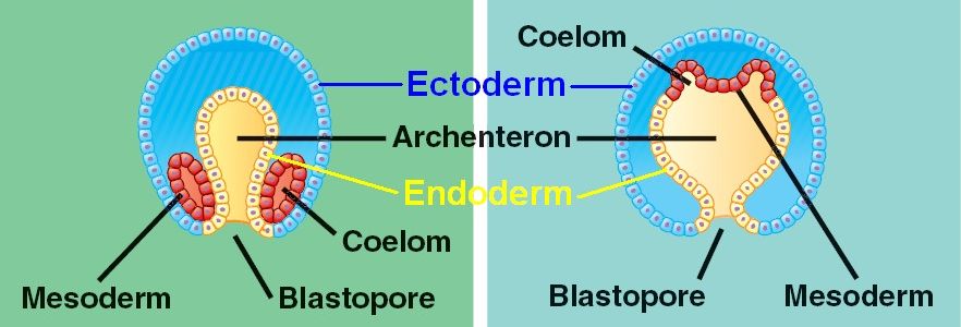

The indention of the blastocoel at the vegetal pole is called the BLASTOPORE. The invaginated region of the vegetal plate is called the ARCHENTERON (or primative gut). After ingression of the primary mesenchyme the vegetal plate invaginates about 1/3 the way into the blastocoel. The cells of the vegetal plate maintain their adhesive interactions with one another and with the hyaline layer. Both these components, fibronectin and the sulfated glycoproteins, are important for the ingression and migratory behavior of the primary mesenchyme.Īfter a period of migratory exploration the primary mesenchyme cells settle into a ring in the vegetal region of the blastocoel. The primary mesenchyme cells synthesize some as yet unidentified surface sulfated glycoproteins. The affinity of the primary mesenchyme cells for fibronectin increases dramatically at the time of ingression. An important component of the basal lamina is fibronectin.

They adhered 100 times more strongly to the basal lamina than to the hyaline layer or other blastomeres.

The migratory primary mesenchyme cells showed just the opposite behavior. Results showed that pre-migratory micromeres adhered 100 times stronger to the hyaline layer and other blastomeres than to the basal lamina. They then added 16 cell stage micromeres, migratory stage primary mesenchyme cells, gastrula ectoderm and endoderm. They first made test plates to which they bound hyaline, test cell monolayers, or basal lamina. This model that suggests differing adhesive interactions is responsible for the ingression of the primary mesenchyme was directly tested by Fink and McClay. These cells first migrate randomly in the blastocoel, but soon become localized on the ventral surface of the blastocoel, where they fuse to form the syncytial cables that will give rise to the calcium carbonate spicules. They are fated to give rise to the SKELETAL RODS of the PLUTEUS LAVA. Cells which are the first to INGRESS are called the PRIMARY MESENCHYME and are derived from the MICROMERES. The cells INGRESS as individuals and not as a sheet (not epiboly), not an INVAGINATION, not an INVOLUTION, not a DELAMINATION. The cells loose their adhesion with the surrounding cells of the blastoderm and are pulled inward into the blastocoel by their filopodial processes. They are also attached at their basal surfaces to the basal lamina.Ĭentrally located cells begin to extend filopodial processes from their inner surface. The blastomeres adhere strongly to the hyaline layer and to each other. The blastula is surrounded by the hyaline layer and the blastomeres have secreted a basal lamina surrounding the blastocoel.

About 24 hrs after hatching of the ciliated blastula the vegetal side of the blastula begins to flatten to form the VEGETAL PLATE.
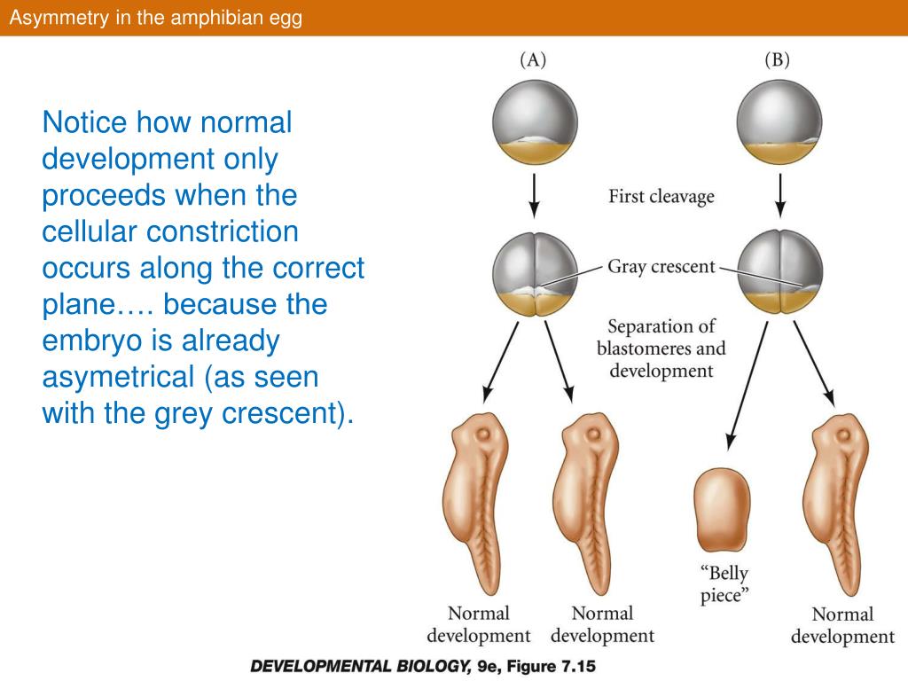
#GREY CRESCENT IN HUMANS BLASTOPORE MOVIE#
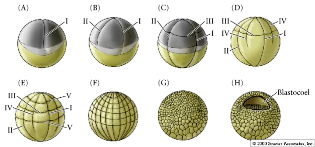
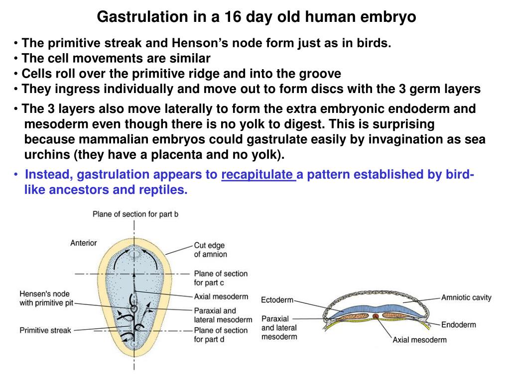
The fate of blastomeres in the hatched blastula can be easily recognized as arising from the early pattern of cleavages and suggests little cell movement during the blastula stage.Įxcellent movie of sea urchin gastrulation from Rachel Fink's "A Dozen Eggs". Remember at the 4th cleavage the animal pole mesomeres divide in the meridinal plane while the vegetal pole cells divide in the equatorial plane asymmetrically to give rise to the 4 macromeres and the 4 micromeres. Even so, blastomeres do not always cleave symmetrically. The egg is isolecithal and cleavage symmetry is radial holoblastic. Remember the cleavage pattern of sea urchins. The same blastomeres end up with the same egg cytoplasm and in the same positions in the blastuala. Cells can migrate as individuals, eg., ingression, or as part of a unit with other cells, eg., invagination, involution, delamination, and epiboly.Ī fate map can be drawn on the egg because of the stereotyped pattern of cleavages. New positions and neighbor relationships are determined by the pattern of cell movements at gastrulation. The initial positions and neighbors of the blastomeres is determined by the pattern of cleavages. Gastrulation is the process of cell movements that give rise to the primary germ layers of the embryo.


 0 kommentar(er)
